SERVICES
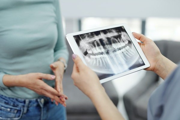
Digital X-Rays
Intraoral periapical radiographs, commonly known as dental X-rays, are one of the most commonly used diagnostic tools in dentistry. They provide detailed images of the teeth, surrounding bone, and supporting structures that cannot be seen with the naked eye. In this article, we will discuss what intraoral periapical radiographs are, how they are used, and their benefits and limitations.
What are intraoral periapical radiographs?
Intraoral periapical radiographs are a type of dental X-ray that is taken from within the mouth. They capture images of individual teeth from the crown to the root, and a small portion of the surrounding bone. These images provide dentists with valuable information about the condition of a tooth and the surrounding structures.
How are intraoral periapical radiographs taken?
Intraoral periapical radiographs are taken using a dental X-ray machine. The patient is positioned in a chair, and the X-ray machine is positioned close to the area of the mouth that needs to be imaged. The dentist or dental assistant will then place a small, flat, plastic device called a “film holder” or “sensor” into the patient’s mouth. The patient will be asked to bite down gently on the device while the X-ray is taken. This process is repeated for each tooth or area that needs to be imaged.
Benefits of intraoral periapical radiographs
Intraoral periapical radiographs provide dentists with valuable information about the teeth and surrounding structures that cannot be obtained through a visual examination. Some of the benefits of intraoral periapical radiographs include:
1. Early detection of dental problems: Intraoral periapical radiographs can detect dental problems such as cavities, tooth decay, and gum disease in their early stages. This allows dentists to provide prompt treatment, which can help prevent further damage to the teeth and surrounding structures.
2. Accurate diagnosis: Intraoral periapical radiographs can provide dentists with a more accurate diagnosis of dental problems. The images can reveal the extent of damage or decay to a tooth and help dentists determine the best treatment plan.
3. Planning for dental procedures: Intraoral periapical radiographs are often used to plan for dental procedures such as root canals, extractions, and dental implants. The images can help dentists determine the location and size of the affected area, which can help them plan the procedure more effectively.
4. Tracking changes over time: Intraoral periapical radiographs can be used to track changes in the teeth and surrounding structures over time. This allows dentists to monitor the progress of dental problems and adjust treatment plans as needed.
Limitations of intraoral periapical radiographs
While intraoral periapical radiographs are a valuable diagnostic tool, they do have some limitations. Some of the limitations of intraoral periapical radiographs include:
1. Radiation exposure: Intraoral periapical radiographs involve exposure to radiation, which can be harmful in large doses. However, the amount of radiation exposure from a single X-ray is minimal and is considered safe for most patients.
2. Limited field of view: Intraoral periapical radiographs capture images of individual teeth and a small portion of the surrounding bone. They do not provide a full view of the mouth, which can make it difficult to detect problems in other areas.
3. Difficulty capturing images: In some cases, it can be difficult to capture clear images using intraoral periapical radiographs. This can be due to factors such as patient movement, the position of the tooth, or the size of the mouth.
4. Inability to capture soft tissue images: Intraoral periapical radiographs are only able to capture

